3D Artificial Intelligence MRI Assessment: The Future of Medical Imaging?
Medical imaging technology has come a long way in the past few decades. One of the latest advancements in this field is 3D artificial intelligence magnetic resonance imaging assessment. This diagnostic imaging allows doctors to get a more accurate picture of what is happening inside a patient’s body. It can be used for both diagnosis and treatment planning. In this blog post, we will discuss the benefits of Springbok 3D artificial intelligence in medical imaging analysis and how it is changing the field of diagnostic imaging.
One of the biggest advantages of artificial intelligence in medical imaging is that it can help to improve accuracy and efficiency. In traditional two-dimensional MRI scans, doctors have to examine a lot of data to get a clear picture of what is happening inside the patient’s body. With the help of artificial intelligence, they can now analyze three -dimensional images which makes the process much more efficient. In addition, artificial intelligence can help to identify lesions and other abnormalities that may be difficult to see with the naked eye.
Diagnostic Medical Imaging
Magnetic resonance imaging technology is getting better and better. This means that we can learn more about muscles, tendons, and bones than ever before. While this data has great value in identifying and diagnosing a number of musculoskeletal abnormalities, only highly sub-specialized radiologists are able to interpret this data with high accuracy. These very real barriers – the significant amount of time required for manual analysis and years of training needed to attain proficiency – prevent clinicians from leveraging the full capabilities of musculoskeletal MRI analysis in any kind of scalable clinical workflow. This challenge is exactly why AI-based imaging analysis is revolutionizing the field. Artificial intelligence-based analytical algorithms can give providers a wide range of experience objective, precise data expeditiously and at scale.
Going Beyond the Scan: How Artificial Intelligence Can Help with Diagnosis
The use of MRI is a growing and incredibly effective method to diagnose musculoskeletal injuries. Many techniques that can help unlock the potential of detailed medical images are supported by scientific studies. These techniques usually involve classifying whether or not a pathology is present. Yet what is missing from some of these techniques is the ability to precisely characterize a musculoskeletal abnormality. MRIs hold great value when they can account for additional factors such as tear location, configuration, size, and the state of an injured ligament, tendon, or meniscus, something that is critical for significant clinical decisions, including electing surgery and understanding outcomes.
For musculoskeletal MRI interpretation to add more value in clinical practice, innovations for determining an abnormality’s presence or absence must be enhanced to include:
- Characteristics and extent of an abnormality.
- Predict related injuries.
- Enforce treatment selection.
- Deliver accurate prognosis.
Improving Patient Outcomes
Diagnostic accuracy is step one to appropriate treatment plan — as misdiagnosis or underdiagnosis can not only derail progress but also exacerbate an issue. Due to limitations in technology, it has not always been possible or feasible to obtain a big picture, yet detailed data for an injured area and its surrounding structures.
By contrast, artificial intelligence in medical imaging diagnostic tools are yielding far more accurate and detailed imaging results — faster and at scale. Efficiencies gained by AI-based data analysis are improving clinical practice.
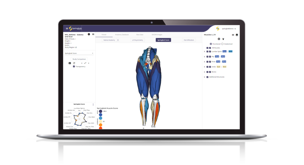
Artificial Intelligence in Medical Imaging of Athletes
As shown in Skeletal Radiology’s review of deep-learning imaging tools, artificial intelligence offers tremendous potential for the field of sports medicine. In addition to streamlining and improving current image-based diagnostics, AI algorithms will continually learn more about human physiology to give scientists, clinicians, and providers even more insight into the human body. The data produced will further improve our understanding of muscle segmentation, complementarity, and optimal function, even with the effects of aging and injury.
AI Techniques as a Tool for Clearer Tissue Imaging and Diagnosis
AI-powered magnetic resonance imaging analysis tools can greatly assist providers in a wide range of clinical decision-making instances. By minimizing the margin of error and generating advanced details about the affected musculoskeletal structure, these technologies allow them to create a truly personalized and progressive care plan. As importantly, the rapid analysis makes this process much more efficient and scalable than traditional MRI-based diagnostic approaches.
The Evolution of How We Analyze Muscle in Sports Medicine
It’s not always easy to track muscle development in athletes — or atrophy in aging or injured individuals. There are a variety of tools and assessments available to measure muscle strengths and weaknesses ranging from rudimentary assessments to advanced imaging analysis. As imaging analysis becomes more sophisticated, we are able to quantify and get deeper insights into muscle health than ever before. These advancements enable us to better understand the key factors in hypertrophy (growth) and atrophy (decline), and most importantly, enable athletes at every level themselves to visualize and understand their muscle health in unprecedented ways.
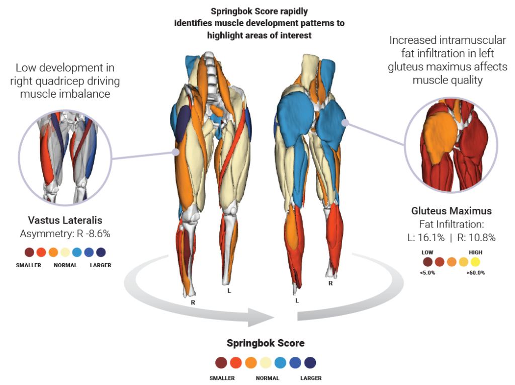
Springbok provides holistic, objective data analysis about musculature, giving health & wellness providers complete confidence when making essential decisions to reduce the risk of injury, rehab smarter, and optimize performance. By acquiring and transforming holistic musculoskeletal MRI data into an interactive 3D model, Springbok’s revolutionary AI-driven technology platform enables true precision medicine.
Data-driven decisions are becoming increasingly common, and essential, in healthcare’s transformation to precision medicine. From personalized strength and conditioning training to customized care and rehab plans based on a person’s individual physiology, acquiring precise data is the key. This is especially true for athletes and their trainers, physical therapists, strength & conditioning coaches, and sports medicine physicians. Whether developing a training program to reduce injury risk, doing a post-surgery rehabilitation assessment or making a critical return to play determination, making these informed decisions require obtaining as much and as accurate information as possible.
Who would benefit from AI-assisted Medical Image Analysis, and why?
Rotator Cuff Tear Repairs
One area where AI algorithms can be very helpful is rotator cuff tears. The shoulder is the second most common site of joint pain and the second most common referral for a joint MRI. Within that population, rotator cuff tears are the most common shoulder injury. Magnetic resonance imaging provides an excellent non-invasive assessment for these tears and can provide data on a number of characteristics known to influence the degree of success of surgery to relieve pain and retain or restore shoulder mobility.
When making the surgical decision, physicians will consider a number of factors, including two important characteristics from the MRI scan – the grade of muscle bulk atrophy and fatty infiltration. These two metrics are essential to the decision-making process as well as the prognosis.
This is because the preoperative degree of atrophy and fatty infiltration of the rotator cuff muscles are predictors that impact function post-operatively. 3D AI MRI algorithms can help PTs, rehab specialists, and physicians develop a patient-specific treatment plan, based on their medical history and unique musculature.
ACL Rehabilitation
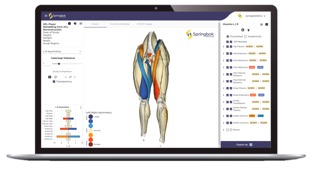
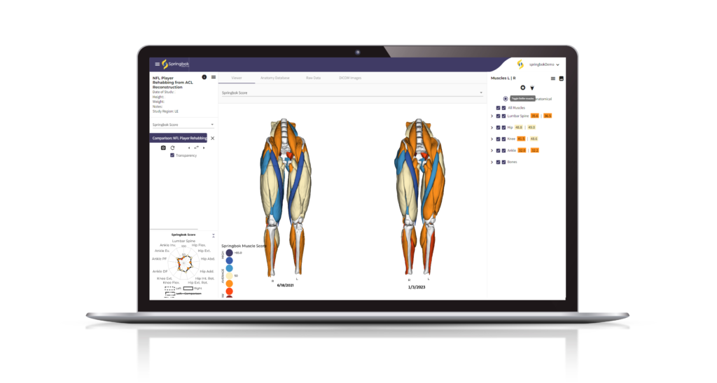
An athlete who sustained an anterior cruciate ligament tear (ACL) on their knee underwent ACL reconstruction and began a rehab regimen to regain strength and return to play. The athlete and their care team needed a faster way to get objective results on recovery progress than traditional assessment tools provide. Clinicians can utilize Springbok analyses post-operatively to:
- Quantify muscle imbalances and development patterns
- Get a holistic view of their unique musculature
- Track changes in response to the rehab conditioning
- Create a road map for optimal recovery
With artificial intelligence-driven MRI assessment, we are able to get an even deeper understanding of muscle health and function. This technology allows us to detect injury at its earliest stages, improve the accuracy of diagnoses, and create more individualized care plans. As we continue to evolve our understanding of human physiology and musculoskeletal structure, artificial intelligence will be essential in helping us make data-driven decisions that improve the health and performance of athletes at all levels.
References
- Skeletal Radiology, “Deep Learning Imaging Tools for Musculoskeletal Medicine”
- Springbok, “How We Analyze Muscle in Sports Medicine”
- Precision medicine, “Data Driven Healthcare”
Have you been injured at some point in your journey?
Are you not achieving your highest level of function?
We’ve helped hundreds of people at all walks in life
get back to performing their best painfree!
MOTUS Client
Janelly Farias
PT's to the Pro's, Stars and Skeptics
Discover why the pro's, protective parents, and weekend warriors trust our 4P Joint By Joint Approach™.

Lorem Ipsum
Daughter
Lorem ipsum dolor sit amet, consectetur adipiscing elit. Nam eget sollicitudin metus, sed euismod purus. Donec quis mauris in ligula luctus accumsan vitae a sem. Morbi hendrerit, justo auctor mattis vulputate, diam sem imperdiet metus, non vehicula arcu arcu in dui. Cras nec sollicitudin diam. Pellentesque finibus metus at dui gravida semper.

Lorem Ipsum
Professional Golfer
Lorem ipsum dolor sit amet, consectetur adipiscing elit. Nam eget sollicitudin metus, sed euismod purus. Donec quis mauris in ligula luctus accumsan vitae a sem. Morbi hendrerit, justo auctor mattis vulputate, diam sem imperdiet metus, non vehicula arcu arcu in dui. Cras nec sollicitudin diam. Pellentesque finibus metus at dui gravida semper.

Lorem Ipsum
Lawyer
Lorem ipsum dolor sit amet, consectetur adipiscing elit. Nam eget sollicitudin metus, sed euismod purus. Donec quis mauris in ligula luctus accumsan vitae a sem. Morbi hendrerit, justo auctor mattis vulputate, diam sem imperdiet metus, non vehicula arcu arcu in dui. Cras nec sollicitudin diam. Pellentesque finibus metus at dui gravida semper.

Lorem Ipsum
NFL
Lorem ipsum dolor sit amet, consectetur adipiscing elit. Nam eget sollicitudin metus, sed euismod purus. Donec quis mauris in ligula luctus accumsan vitae a sem. Morbi hendrerit, justo auctor mattis vulputate, diam sem imperdiet metus, non vehicula arcu arcu in dui. Cras nec sollicitudin diam. Pellentesque finibus metus at dui gravida semper.

Lorem Ipsum
NFL
Lorem ipsum dolor sit amet, consectetur adipiscing elit. Nam eget sollicitudin metus, sed euismod purus. Donec quis mauris in ligula luctus accumsan vitae a sem. Morbi hendrerit, justo auctor mattis vulputate, diam sem imperdiet metus, non vehicula arcu arcu in dui. Cras nec sollicitudin diam. Pellentesque finibus metus at dui gravida semper.
It's time to toss out the old school
physical therapy playbook.
We'll never hand you a stack of those black and white exercise printouts.
MOTUS Client
Sam Darnold
MOTUS Client
Kyle Allen
Level up on rehab and prevention and get back to the activities you love
Schedule A Call
We'll walk you through our 4P Joint Approach™ and set up your 60-minute 1:1 consultation.
Get Your Personalized 4P Plan
We'll pinpoint the source of your pain and design a plan to restore movement along the entire kinetic chain.
Start Moving Again
Get an edge on injury prevention, relieve joint and muscle pain, and return to activities you love with confidence.
Stop wondering if you'll ever
get back to being you.

Professional Photographer
3 Ways to Level Up Your Rehab and Injury Prevention With Us

